
The Obstructed Kidney
in the Fetus, Infant, and Child
by
The kidney can become obstructed…
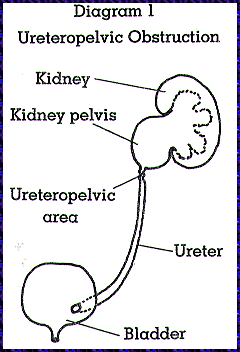
when there is interference with the flow of urine from the kidney to the bladder. The result is hydronephrosis, or swelling of the kidney. In infants and children, this is usually a congenital condition (they are born with it). Because of the frequent use of ultrasound examination in current obstetrical practice, congenital urinary tract obstruction is often diagnosed early, since it can be seen at 15 weeks of fetal development. Fetal hydronephrosis is seen with ultrasound in approximately one of every 600-800 fetuses examined. The most common area of kidney blockage in the fetus or infant is at the ureteropelvic junction where urine drains from the kidney to the ureter (Diagram 1).

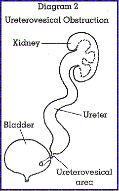
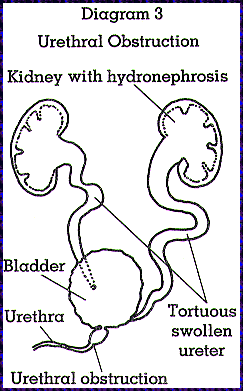
Less common are cases where the blockage occurs at the ureterovesical junction, where urine drains from the ureter to the bladder (Diagram 2), or where obstruction occurs at the urethra (Diagram 3). Urethral obstruction usually causes hydronephrosis (swelling) in both kidneys.
Obstruction can be diagnosed… with different methods, depending upon the age of the fetus, infant or child. When the fetus is in utero, serial ultrasound is used to see any progression in swelling of the kidney. Sound waves are passed through the mother’s abdomen and uterus to the fetal kidneys, which can then be seen. In almost all cases, fetal ultrasound identifies kidney swelling and other lesions of the urinary tract, which allows prompt evaluation at birth. Mild kidney swelling may disappear after birth, however such disappearance is rare with more severe swelling or hydronephrosis.
The infant or child is also examined with ultrasound as well as with a renal scan called a Lasix renogram. For this test, a small amount of material (isotope) is injected into the child’s vein to see how long it takes to enter and leave the kidney. Analysis of the results helps to define the degree of obstruction in the kidney and to evaluate the amount of function in each of the kidneys. Another examination often performed when evaluating an infant or child with hydronephrosis is a voiding cystourethrogram (VCUG). A small tube is placed into the infant or child’s urethra and the bladder is filled with fluid visible on an x-ray. All of these tests can be performed on an outpatient basis with the infant or child awake.
Treatment is recommended… to prevent loss of function in the blocked kidney. An obstructed kidney is susceptible to urinary tract infection which can further destroy its function as well as cause pain and bleeding. The best time for relief of the obstruction depends on four factors: the degree of obstruction, any loss of kidney function, possible urinary infection, and the location of the blockage in the urinary tract. Fetal surgery is considered only in extremely rare circumstances when both kidneys are severely affected. This is currently a form of experimental surgery. For newborns with renal obstruction, there is much clinical evidence that early relief of blockage after birth encourages the return of the most amount of function in the blocked kidney. Particularly in the most common cases (blockage at the ureteropelvic area), early relief within the first few months of life preserves kidney tissue and encourages the greatest return of function. Early relief of blockage can also allow the previously obstructed kidney to grow before the other kidney takes over some of its function (compensatory hypertrophy). With appropriate anesthesia in generally healthy infants, surgery is safe and usually successful. The older infant or child with obstruction should also be surgically treated to stabilize the kidney, prevent urinary tract infection or any further loss of renal function, and hopefully, allow recovery of function.
Surgical treatment of obstructions… involves removal of the narrowed or poorly functioning area causing the blockage. At the ureteropelvic or ureterovesical areas, this calls for a surgical incision through the side or lower abdomen with reattachment of the ureter to the kidney or bladder as necessary. Sometimes an enlarged and swollen ureter must also be narrowed as part of the repair. The operation requires two to three hours to perform and is successful in 95% of the cases. Treatment of urethral obstruction varies depending upon the severity of the blockage. This discussion serves as an introduction to the problem of the obstructed kidney. I encourage you to discuss with me your questions and concerns about this condition and the procedures used to correct it.
Return to the PedsUroLogic Home Page

Richard M. Parker, M.D., F.A.A.P., F.A.C.S.
[email protected]
Arnold Medical Pavilion
1221 Madison Street
Suite 412
Seattle, WA 98104 USA
Email:[email protected]
The Olympic Center
1310 116th Avenue N.E.
Suite B
Bellevue, WA 98004 USA
Email:[email protected]